
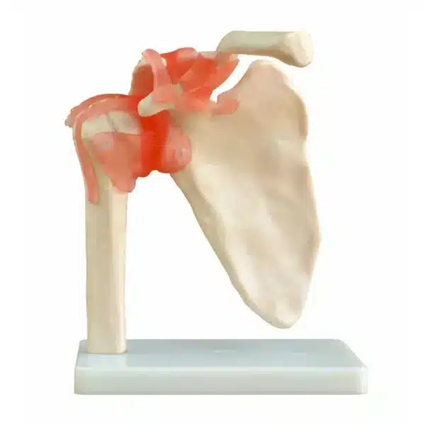
Shoulder Model
Discover how this Shoulder Model transforms abstract anatomical knowledge into tangible, interactive learning.
This Shoulder Model is an essential educational tool used in medical, anatomical, and physiotherapy training to demonstrate the intricate complexity of the human shoulder joint. This life-sized or scaled model accurately replicates the bones, ligaments, tendons, and sometimes musculature of the glenohumeral joint—the body’s most mobile and consequently unstable ball-and-socket articulation.
A high-quality teaching model typically features detachable components, allowing for hands-on exploration. The bony structures—including the clavicle, scapula, and proximal humerus—are often color-coded. Key anatomical landmarks, such as the acromion, coracoid process, and glenoid fossa, are clearly defined. Many models include a transparent overlay of the joint capsule or a cut-away section to reveal the internal architecture.
The model’s primary pedagogical value lies in its ability to illustrate dynamic function. Students can visualize the relationship between the humeral head and the shallow glenoid cavity, understanding the critical stabilizing role of the labrum and ligaments like the coracohumeral and glenohumeral ligaments. Models incorporating rotator cuff muscles (supraspinatus, infraspinatus, teres minor, subscapularis) demonstrate their concerted action in securing the joint and enabling a wide range of motion.
In clinical instruction, the model is indispensable for explaining common pathologies. Educators can point to areas prone to injury, such as the site of rotator cuff tears, the mechanism of anterior dislocation, or the impingement of subacromial structures. It provides a three-dimensional reference far superior to textbook diagrams, fostering spatial understanding crucial for surgical planning, physical examination techniques, and rehabilitation strategies.
Ultimately, this Shoulder Model transforms abstract anatomical knowledge into tangible, interactive learning. It bridges the gap between two-dimensional theory and the living body, providing an invaluable foundation for future healthcare professionals to diagnose, treat, and understand shoulder dysfunction.
Q & A
 Shoulder Model
Table of Contents
Frequently Asked Questions
What specific anatomical structures does this shoulder model show, and how does it demonstrate shoulder instability?
This life-sized model meticulously replicates the core structures of the glenohumeral joint. You will observe the bony architecture—the scapula, clavicle, and humerus—with key landmarks like the glenoid fossa and acromion clearly defined. Critically, it showcases the soft tissues that provide stability: the labrum deepening the socket, and ligaments such as the coracohumeral and glenohumeral ligaments that reinforce the capsule.
The model’s pedagogical strength lies in illustrating why this mobility comes at a cost. You can physically see the dramatic size discrepancy between the large humeral head and the shallow glenoid cavity, visually explaining the joint’s inherent instability. By detaching components, you can appreciate how the surrounding ligaments and, in advanced models, the rotator cuff musculature, are essential to secure the joint, preventing dislocation during movement.
How can this shoulder model be used to understand common shoulder injuries and clinical procedures?
This model is an indispensable clinical teaching tool for translating anatomy into practice. Instructors use it to pinpoint common injury sites, such as the vulnerable area of the supraspinatus tendon in rotator cuff tears, or the anterior-inferior labrum in Bankart lesions following dislocation. The three-dimensional perspective is crucial for understanding impingement syndromes, as one can visualize how structures become compressed under the acromion.
Furthermore, this shoulder model provides a tangible reference for procedural planning and education. Students can trace paths for therapeutic injections, understand the spatial relationships critical for surgical approaches, and practice physical examination maneuvers on a static structure before applying them to patients. This bridges theoretical knowledge directly to clinical assessment and intervention strategies.
Is this shoulder model designed for hands-on interaction, and what are its key features for education?
Absolutely. High-quality educational models are explicitly designed for interactive, hands-on exploration. A key feature is the detachable components, allowing users to disassemble the joint to study individual bones and their articulating surfaces. Many models include transparent sections or overlay windows to reveal the internal arrangement of ligaments and the joint capsule without full disassembly.
Additional educational features include color-coding on bony structures and muscular attachments for immediate identification, as well as realistic ligamentous tension that permits demonstration of normal and excessive joint play. This interactivity transforms passive learning into an active experience, fostering a deeper spatial understanding of shoulder biomechanics, muscle function, and the relationship between form and function that flat images cannot provide.


Reviews
There are no reviews yet