
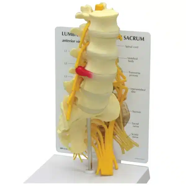
Vertebrae with Sacrum Model
Learn why this Vertebrae with Sacrum Model is an indispensable resource for students and professionals in anatomy, chiropractic, physical therapy, and medicine.
This meticulously detailed Vertebrae with Sacrum Model is an essential educational tool, offering a three-dimensional exploration of the lower spine’s complex architecture. The model typically presents a life-sized or slightly enlarged articulation, encompassing the five lumbar vertebrae (L1-L5), the sacrum, and the coccyx. Each bony element is accurately shaped to demonstrate the unique anatomical features critical to both structure and function.
The lumbar vertebrae are clearly distinguished by their large, kidney-shaped vertebral bodies, designed to bear the majority of the body’s weight. Their short, blunt spinous processes project horizontally, while the sturdy transverse processes extend laterally. Between each vertebral body, removable discs representing the intervertebral fibrocartilage illustrate their role as shock absorbers and facilitators of limited movement.
The centerpiece of the Vertebrae with Sacrum Model is the sacrum, a fused, triangular bone forming the posterior wall of the pelvis. The model highlights its five fused segments, the superior articular processes that articulate with L5, and the prominent sacral promontory. The dorsal surface displays the median sacral crest, posterior sacral foramina, and the sacral hiatus. The lateral auricular surfaces, crucial for the sacroiliac joints, are also evident. The model concludes with the small, segmented coccyx, or tailbone, attaching inferiorly to the sacrum.
Key ligaments, such as the anterior and posterior longitudinal ligaments, are often represented in flexible material, showing their role in stabilizing the spinal column. The spinal canal, formed by the alignment of vertebral foramina, is visibly patent, protecting the neural structures within.
This dynamic Vertebrae with Sacrum Model allows for demonstration of spinal flexion, extension, and lateral bending, providing insight into the biomechanics of the lower back and lumbosacral junction. It is an indispensable resource for students and professionals in anatomy, chiropractic, physical therapy, and medicine, transforming abstract textbook descriptions into a tangible, interactive understanding of spinal anatomy, common injury sites, and clinical significance.
Q & A
 Vertebrae with Sacrum Model
Table of Contents
Frequently Asked Questions
What specific educational value does this vertebrae with sacrum model offer compared to a standard textbook image?
This model transforms two-dimensional, static information into a tangible, three-dimensional learning experience. A textbook can describe the sacrum’s shape, but holding the solid, triangular bone reveals its true role as the keystone of the pelvic girdle. You can physically trace the path of nerves through the intervertebral and sacral foramina, understanding impingement risks in a way flat diagrams cannot convey.
Furthermore, its articulating design allows for dynamic demonstration. You can manually simulate spinal movements like flexion and extension, observing how the facets glide and how the ligaments would tension. This hands-on interaction is crucial for grasping biomechanics, common injury mechanisms, and the clinical rationale behind treatment modalities, making it indispensable for tactile learners in clinical fields.
How does this vertebrae with sacrum model illustrate the transition from mobility to stability in the lumbosacral region?
The model expertly visualizes this critical anatomical transition. The five separate lumbar vertebrae, with their removable discs and flexible ligaments, clearly represent the mobile segment designed for weight-bearing and movement. This contrasts sharply with the singular, fused mass of the sacrum below, embodying structural stability.
The junction itself, the lumbosacral joint (L5-S1), is a focal point of study. The model shows how L5’s massive body articulates with the slanted base of the sacrum at the promontory, creating a sheer line that is a common site for spondylolisthesis. Seeing this angle in 3D clarifies why this joint withstands immense shear forces and is frequently associated with degeneration and pain, highlighting the body’s compromise between flexibility and robust support.
What clinical conditions or structures can this model help to explain?
This vertebrae with sacrum model serves as a vital visual aid for numerous clinical concepts. It clearly identifies vulnerable areas: the intervertebral discs for herniations, the facet joints for arthritis, and the L5-S1 junction for instability. The open spinal canal and nerve exit foramina allow educators to demonstrate how stenosis or a bulging disc can compress spinal nerves, leading to symptoms like sciatica.
Furthermore, it details pelvic anatomy relevant to injections and pain management. The model’s accurate depiction of the sacral hiatus provides a perfect guide for teaching caudal epidural procedures. Similarly, the auricular surfaces demonstrate the sacroiliac joints, often a source of referred pain. By offering a concrete reference, this vertebrae with sacrum model bridges foundational anatomy to patient assessment and treatment planning.


Reviews
There are no reviews yet