
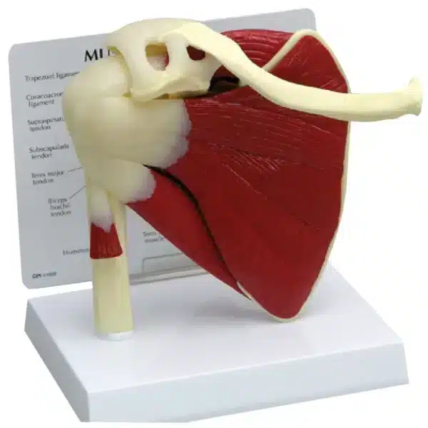
Muscled Shoulder Model
Discover how this muscled shoulder model helps to bridge the gap between theoretical knowledge and clinical understanding.
A muscled shoulder model is an essential anatomical teaching tool that vividly illustrates the complex structure and mechanics of the human shoulder girdle. Typically life-sized, this specialized model dissects away the skin to reveal the intricate layers of musculature, tendons, and often underlying bony landmarks like the clavicle, scapula, and proximal humerus.
The primary didactic value lies in its three-dimensional, hands-on nature. Students can visually trace the origin, insertion, and fiber direction of each major muscle—from the superficial deltoid and trapezius to the deeper rotator cuff muscles (supraspinatus, infraspinatus, teres minor, and subscapularis). This clarity is impossible to achieve with textbook diagrams alone. Many models are partially detachable or color-coded, allowing for the study of individual muscles and their relationship to the joint capsule and bony prominences.
Educators use this model to teach fundamental concepts of shoulder function, including the coordination required for abduction, rotation, and the stability provided by the rotator cuff. It demonstrates how muscular forces translate into the wide range of motion characteristic of this ball-and-socket joint, while also highlighting vulnerable points for common injuries like impingement or rotator cuff tears.
By providing a tangible, detailed reference, the muscled shoulder model bridges the gap between theoretical knowledge and clinical understanding. It is indispensable in classrooms for medical, physical therapy, kinesiology, and athletic training students, fostering a deeper comprehension of one of the body’s most mobile and clinically significant joints.
Q & A
 Muscled Shoulder Model
Table of Contents
Frequently Asked Questions
What specific anatomical structures does a typical muscled shoulder model show, and how is it used for teaching?
A comprehensive muscled shoulder model meticulously details the entire shoulder girdle. It presents the bony framework—the clavicle, scapula, and humerus—serving as the anchor points. Superficially, it displays major muscles like the deltoid and trapezius, which are often removable. Beneath these, the model reveals the deep, critical layer of the rotator cuff muscles (supraspinatus, infraspinatus, teres minor, subscapularis), along with associated tendons and the joint capsule.
Educators leverage this three-dimensionality to move beyond static diagrams. Students can physically trace a muscle’s path from its origin to insertion, directly observing how fiber direction dictates function. This hands-on interaction is pivotal for teaching complex concepts like the coordinated mechanics of abduction or the stabilizing role of the rotator cuff during movement, making abstract principles tangibly clear.
How does this muscled shoulder model help in understanding shoulder injuries and clinical conditions?
The model serves as a vital bridge to clinical application by visually isolating vulnerable anatomical relationships. For instance, it clearly demonstrates how the supraspinatus tendon passes beneath the acromion, providing an immediate visual for understanding subacromial impingement syndrome. The attachment points of the rotator cuff tendons on the humeral head vividly illustrate how tears occur and why they affect specific motions.
Furthermore, by seeing the close proximity of muscles, tendons, and bursae, students can better comprehend pathologies like tendonitis, bursitis, and instability. The model allows for the simulation of injury mechanisms, such as how a dislocation might occur, fostering a predictive understanding of which structures are at risk. This direct visualization is foundational for diagnosing and planning rehabilitation for common shoulder dysfunctions.
What features should I look for when selecting a muscled shoulder model for educational or professional use?
Key features enhance a model’s didactic value. Prioritize anatomical accuracy and high-quality materials that withstand frequent handling. Color-coding and detachable components—especially a removable deltoid and rotator cuff muscle group—are essential for studying deep structures. Some advanced models include labeled ligamentous structures or a transparent overlay showing range of motion.
Consider the educational context. A basic, durable model suffices for introductory anatomy, while a detailed, dissectible model is crucial for medical or physical therapy training. The optimal model balances detail with clarity, avoiding visual clutter, to effectively illustrate the dynamic interplay between form and function that defines this complex joint.


Reviews
There are no reviews yet