
Anatomical Spine Model
Discover why this Anatomical Spine Model is an invaluable resource for students and medical professionals, providing a static yet dynamic representation of a dynamic disease process.
This meticulously crafted Anatomical Spine Model serves as an exceptional educational tool, specifically designed to illustrate the intricate and often painful process of degenerative change in the lumbar spine. It transforms a complex, internal physiological progression into a clear, tangible, and hands-on learning experience, ideal for both clinical study and direct patient education.
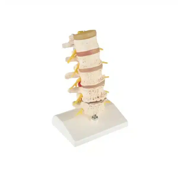
The Anatomical Spine Model’s core strength lies in its detailed depiction of five progressively serious stages of degeneration, charting a clear visual journey from spinal health to significant pathology. Beginning at the top, it presents an intervertebral disc in a healthy, hydrated state paired with a pristine L1 vertebra, establishing a baseline of normal anatomy. From this point, the descent down the model reveals a carefully sequenced pathology. The next level demonstrates the initial bulge or protrusion of a disc, accompanied by an L2 vertebra showing the first osteophytic lipping and bony changes, marking the onset of degeneration.
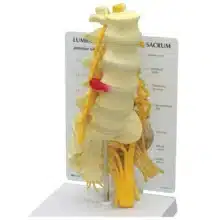
As the progression continues, the conditions become more pronounced. The model shows a medial disc prolapse (herniation) at one level, followed by an L3 vertebra with advanced bony growths and facet joint involvement. The climax of the pathological narrative is powerfully displayed at the lower lumbar segments. Here, the model illustrates a critical intraforaminal disc prolapse on the right, while the accompanying L4 vertebra exhibits severe bony constriction specifically within the left intervertebral foramen. This spatial specificity is crucial for understanding radicular symptoms.
The sequence culminates with an extremely narrowed, desiccated disc and the L5 vertebra, which mirrors the foraminal stenosis seen at L4. This final stage vividly demonstrates how this bony encroachment applies direct pressure on the spinal nerve root (the L5 root on the left), providing an immediate and understandable correlate for patients suffering from sciatica, numbness, or weakness.
Beyond its illustrative power, this Anatomical Spine Model is fully disassemblable. Each vertebra and disc can be separated, allowing for close inspection of individual structures—from vertebral bodies and processes to the annulus fibrosis and nucleus pulposus of each disc. This interactivity deepens anatomical understanding and facilitates targeted explanation.
In summary, this Anatomical Spine Model is an invaluable resource. For students and medical professionals, it provides a static yet dynamic representation of a dynamic disease process. For patients, it demystifies their diagnosis, offering a clear, visual narrative that explains the source of their pain, the rationale behind treatment options, and the importance of preventative spine health. It bridges the gap between textbook diagrams and lived experience, making the invisible both visible and comprehensible.
Q & A
 Anatomical Spine Model
Table of Contents
Frequently Asked Questions
How does this anatomical spine model specifically help in patient consultations?
This spine model serves as a powerful communication bridge, transforming an abstract diagnosis into a tangible, visual story. For a patient struggling to understand their MRI report or the source of their sciatica, pointing to the specific stage of herniation or foraminal stenosis on the model provides immediate clarity. It visually explains how a bulging disc or bony growth directly impinges on a nerve root, making the correlation between their anatomical change and their symptoms—like radiating pain or numbness—instantly understandable.
This demystification is crucial for informed consent and building a therapeutic alliance. By using an anatomical spine model to illustrate the “why” behind a treatment plan, whether it’s physical therapy to create space or a surgical intervention to relieve pressure, patients can visualize their condition and participate more fully in their care decisions. It moves the conversation from a technical description to a shared understanding of their personal pathology.
What are the key educational benefits of this anatomical spine model for students and clinicians?
The model’s core strength is its sequenced, static depiction of a dynamic degenerative process, offering a “timeline in space” that textbooks cannot. It allows learners to visually trace the cascade from a healthy disc to severe stenosis, reinforcing the interconnected anatomy of discs, vertebrae, joints, and nerve pathways. This solidifies understanding of how structural changes directly lead to clinical syndromes.
Furthermore, its fully disassemblable nature provides hands-on, three-dimensional learning. Students can remove each vertebra and disc to inspect the articular facets, vertebral bodies, and the anatomy of the annulus and nucleus. This interactivity deepens spatial comprehension of how a herniation protrudes or how osteophytes form, transforming passive observation into an active exploration that cements complex anatomical and pathological concepts.
Is this anatomical spine model anatomically accurate, and what materials is it made from?
Yes, the model is meticulously crafted for anatomical fidelity, based on a typical lumbar spine (L1-L5) with a sacrum. It accurately depicts vertebral structures, including the spinous and transverse processes, facet joints, and the critical intervertebral foramina where nerve roots exit. The discs show realistic differentiation between the annulus fibrosus and nucleus pulposus at various pathological stages.
Constructed from high-quality, durable synthetic materials, it is designed for repeated handling in clinical and educational settings. The vertebrae are typically made from a rigid, color-fast plastic, while the discs are fashioned from a flexible, resilient material to demonstrate texture and compression. This robust construction ensures the model can withstand frequent disassembly while maintaining the precise detail necessary for effective demonstration and study.


Reviews
There are no reviews yet