
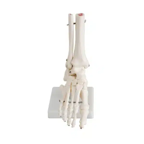
Foot and Ankle Model
Find out how a foot and ankle model can help to demystify the architecture that supports the entire human body.
The human foot and ankle comprise a remarkable biomechanical system of 28 bones, 33 joints, and over a hundred muscles, tendons, and ligaments, all working in concert to provide stability, mobility, and shock absorption. A foot and ankle model serves as an indispensable educational and clinical tool, translating this complexity into a tangible, three-dimensional form for detailed study.
These anatomical models are typically life-sized and crafted from durable materials like high-grade plastics, with bones colour-coded for identification and ligaments, tendons, and neurovascular structures often represented in flexible, distinct colours. The most sophisticated versions are biomechanical, featuring articulated joints that replicate the critical motions of inversion/eversion at the subtalar joint and dorsiflexion/plantarflexion at the ankle. Some foot and ankle models are dissectible, allowing for the removal of layered structures to explore deep anatomy, such as the plantar fascia or the intricacies of the sinus tarsi.
The primary utility of these models spans multiple disciplines. In medical and podiatry schools, they provide students with a hands-on understanding of spatial relationships—how the peroneal tendons wrap around the lateral malleolus, or the convergence of the medial and lateral plantar nerves. For orthopaedic surgeons, they are vital for preoperative planning and patient communication, visually illustrating pathologies like bunions, ankle fractures, or Achilles tendon ruptures. Physical therapists use them to explain rehabilitation protocols, demonstrating the biomechanics of gait and the impact of orthotic interventions.
Ultimately, a foot and ankle model demystifies a foundational yet intricate structure. It moves learning beyond textbook diagrams, offering a static yet dynamic representation of the architecture that supports the entire human body. By providing clarity on form and function, it becomes a cornerstone for education, diagnosis, and effective treatment planning in lower limb health.
Related products: Hand and Wrist Model.
Q & A
 Foot and Ankle Model
Table of Contents
Frequently Asked Questions
What are the key anatomical features and structures that a high-quality foot and ankle model should accurately represent?
A superior model will meticulously replicate the 28 bones, 33 joints, and major soft-tissue structures. Bones should be colour-coded for immediate identification, while critical ligaments (like the deltoid and lateral collateral complexes), tendons (such as the Achilles, tibialis posterior, and peroneals), and neurovascular bundles must be depicted in distinct, flexible materials to show their anatomical courses. Essential biomechanical features include an articulated ankle joint for dorsiflexion/plantarflexion and a subtalar joint that allows for inversion/eversion, mimicking the foot’s natural triplanar motion.
The value of the foot and ankle model is enhanced by layered or dissectible components, enabling the study of deep structures like the plantar fascia, intrinsic foot muscles, and the contents of the tarsal tunnel. Accurate spatial relationships—for instance, how the peroneal tendons pass posterior to the lateral malleolus—are paramount for translating the model into clinical understanding, making it an indispensable tool for visualising complex interactions within a confined anatomical space.
In what professional or educational settings are these foot and ankle models most commonly used, and what specific purposes do they serve?
These models are foundational in academic and clinical environments. In medical, podiatric, and physical therapy schools, they transform two-dimensional textbook knowledge into a tangible, three-dimensional learning experience, allowing students to grasp spatial relationships and biomechanical principles hands-on. For orthopedic surgeons and podiatrists, models are crucial for preoperative planning of complex reconstructions or fracture repairs, and for visually explaining surgical procedures and pathologies—like hallux valgus or ankle instability—to patients in a clear, accessible manner.
Furthermore, in physical therapy and sports medicine clinics, practitioners use these models to demonstrate gait mechanics, injury mechanisms, and the function of therapeutic devices or orthotics. By illustrating how muscles, tendons, and joints work in concert during movement, therapists can effectively justify rehabilitation protocols and foster patient compliance. Thus, the model bridges the gap between theoretical knowledge and practical application across the continuum of care.
What material and design considerations are most important for foot and ankle model durability, accuracy, and educational value?
The ideal foot and ankle model construction employs high-grade, durable plastics or resins for bone structures to withstand frequent handling, while ligaments and tendons are best made from flexible, colour-contrasting polymers to demonstrate their tensile nature without tearing. Biomechanical accuracy is paramount; joints must have realistic articulation to simulate true physiological ranges of motion, not just static positioning. A life-sized scale is essential for appreciating true anatomical proportions and for clinical correlation.
Educational value is significantly enhanced by strategic design choices. Colour-coding is standard for quick identification, but advanced models offer dissectible layers—allowing removal of the plantar fascia, for example, to reveal deeper musculature—or interchangeable parts showing pathological conditions. The most effective models strike a balance between detailed complexity and clear, uncluttered representation, ensuring they elucidate rather than overwhelm the user, thereby serving as a lasting reference for anatomy and biomechanics.


Reviews
There are no reviews yet