
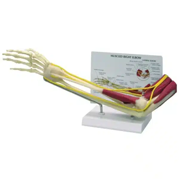
Muscled Elbow Joint Model
Discover how the Muscled Elbow Joint Model presents a comprehensive study of the right elbow joint.
This detailed, life-sized anatomical Muscled Elbow Joint Model presents a comprehensive study of the right elbow joint, meticulously crafted to illustrate the complex interplay between its muscular, skeletal, and connective tissues. Measuring 22″ x 10″ x 5″, the model offers an immersive, true-to-scale view, anchored on a stable plastic base for clear display and study.
Central to the Muscled Elbow Joint Model are the three primary bones of the articulation: the distal end of the humerus, the proximal ulna with its prominent olecranon process, and the proximal radius. These bones form the foundational hinge and pivot joints that allow for flexion, extension, and rotation of the forearm. Attaching to and moving these bones are the major muscle groups of the upper arm and forearm, including the biceps brachii, triceps brachii, brachialis, and the forearm extensors and flexors, showcasing their precise origins and insertions.
Critical for joint stability, the model accurately details the ligaments, such as the ulnar collateral and radial collateral ligaments, which tether the bones together. Accompanying these structures are the key nerves, like the ulnar nerve, depicted in their relative anatomical positions as they traverse the joint area.
Designed for both educational and clinical environments, the Muscled Elbow Joint Model is entirely latex-free for safe handling. It is accompanied by a removable patient education card, which serves as a valuable reference tool to enhance explanation and facilitate understanding of elbow anatomy, common injuries, and biomechanics for students, healthcare professionals, and patients alike.
Q & A
 Muscled Elbow Joint Model
Table of Contents
Frequently Asked Questions
What specific educational or clinical applications is this muscled elbow joint model best suited for?
This model is an exceptional multidisciplinary tool designed for both in-depth study and clear patient communication. In academic settings, such as medical, physical therapy, or sports science programs, it allows students to meticulously explore the integration of bones, muscles, and ligaments that enable the elbow’s hinge and rotation. The true-to-life scale and detailed musculature are perfect for understanding biomechanics, common injury mechanisms, and surgical approaches.
In a clinical environment, such as an orthopaedic or physiotherapy office, it becomes an invaluable visual aid. The model helps healthcare professionals explain conditions like tennis elbow (lateral epicondylitis), ligament sprains, fractures, or ulnar nerve entrapment to patients in an accessible, tangible way. The accompanying removable education card further enhances this, allowing for structured explanations of anatomy, injury, and rehabilitation protocols.
How does the design of this muscled elbow joint model enhance the study of anatomy compared to basic bone-only models?
While skeletal models show structure, this muscled model reveals crucial function. It moves beyond static anatomy to demonstrate dynamic physiology. The comprehensive depiction of major muscle groups—like the biceps brachii, triceps, and forearm flexors—with their precise origins and insertions, illustrates exactly how skeletal movement is generated and controlled through contraction.
Furthermore, the inclusion of key ligaments (collateral ligaments) and nerves (ulnar nerve) in their proper spatial relationships is critical. This allows learners to visualize not just movement, but also stability and potential vulnerability. Seeing how the ulnar nerve passes behind the medial epicondyle, for instance, instantly clarifies why a direct impact causes the sensation of “hitting your funny bone.” This layered, integrated approach fosters a superior understanding of the elbow as a complete, working system.



Reviews
There are no reviews yet