
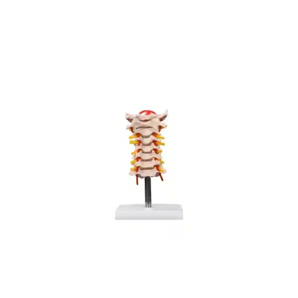
Flexible Cervical Vertebral Column
Discover why the Flexible Cervical Vertebral Column serves as a vital tool for enhancing understanding of spinal health, pathology, and function.
The Flexible Cervical Vertebral Column is a detailed anatomical model designed as an essential teaching aid for clinical, classroom, and hospital settings. Its primary purpose is to demonstrate the complex movement and biomechanical functions of the upper spine. The model accurately represents key anatomical structures, including the occipital plate of the skull, all seven cervical vertebrae, and the intervertebral discs situated between them.
Internally, this flexible cervical vertebral column features critical neural and vascular components. The spinal cord runs through the vertebral foramen, while the cervical nerve roots are shown branching out appropriately. The path of the vertebral arteries, which travel through the transverse foramina to supply blood to the brain, is also clearly illustrated. This comprehensive detail allows educators and medical professionals to explain the relationship between the bony structures, nervous system, and vascular supply in a dynamic, hands-on manner.
Constructed from flexible, latex-free materials, the model is both durable and safe for use in various environments. Its articulated design allows it to be bent and tilted, realistically simulating the normal ranges of motion of the neck—including flexion, extension, lateral bending, and rotation. This functionality makes it ideal for teaching about spinal kinematics, common injuries, and the importance of cervical alignment.
Practical and portable, the Flexible Cervical Vertebral Column measures 7 inches in height and weighs approximately one pound. Its compact size and light weight facilitate easy transport and demonstration at a patient’s bedside, in a lecture hall, or during a clinical consultation. By providing a three-dimensional, manipulable representation of cervical anatomy, the Flexible Cervical Vertebral Column serves as a vital tool for enhancing understanding of spinal health, pathology, and function.
Q & A
 Flexible Cervical Vertebral Column
Table of Contents
Frequently Asked Questions
What specific educational or clinical scenarios is this flexible cervical vertebral column best suited for?
This model is an essential tool across multiple educational and clinical settings. In academic environments, such as university anatomy labs and physical therapy classrooms, it provides students with a three-dimensional, hands-on understanding of cervical anatomy that static textbooks cannot offer. Instructors use it to demonstrate complex concepts like spinal kinematics, the relationship between bony structures and soft tissues, and the biomechanics of normal neck movement.
In clinical contexts, it becomes a powerful patient communication aid. Healthcare professionals, including physicians, chiropractors, and physiotherapists, use the model at the bedside or in consultation rooms to visually explain conditions such as herniated discs, nerve root impingement, cervical stenosis, or the mechanics of whiplash injuries. Its ability to simulate realistic motion helps patients better understand their diagnosis, the nature of their injury, and the rationale behind prescribed treatments or surgical interventions, thereby improving patient education and adherence to care plans.
How does the flexible design of the column enhance learning compared to a static model?
The flexible, articulated design is the core educational advantage of this model, transforming it from a passive display into an active learning tool. By allowing learners to manually perform and observe flexion, extension, lateral bending, and rotation, it creates a kinesthetic understanding of the spine’s range of motion and its limitations. This dynamic demonstration is crucial for comprehending functional anatomy and the principles of biomechanics.
Furthermore, the flexibility allows educators to illustrate pathological conditions and common injury mechanisms in a visceral way. For instance, one can simulate hyperextension to demonstrate whiplash, or show how excessive lateral bending can compress nerve roots or affect the vertebral artery pathway. This interactive capability fosters a deeper comprehension of the close—and often vulnerable—relationship between the moving vertebral column, the delicate spinal cord, the exiting nerves, and the vital vascular structures, which is fundamental for students in neurology, orthopedics, and rehabilitation sciences.
What key anatomical features are included in the flexible cervical vertebral column , and why are they important for a comprehensive understanding?
This model meticulously integrates the critical osteological, neurological, and vascular components of the cervical spine. It includes all seven cervical vertebrae (C1-C7), the occipital bone of the skull, and the intervertebral discs, providing a complete view of the bony architecture and the joints that facilitate movement and absorb shock.
Most importantly, this flexible cervical vertebral column goes beyond bone to show the spinal cord running through the vertebral canal, the paired cervical nerve roots branching from the cord and exiting through the intervertebral foramina, and the vertebral arteries traversing the transverse foramina. This integrated representation is vital for teaching how spinal alignment and movement directly impact the nervous and circulatory systems. Understanding this relationship is essential for diagnosing conditions where bone spurs or disc herniations impinge on nerves (causing radiculopathy) or compromise blood flow to the brain, making the model indispensable for a holistic grasp of cervical spine function and dysfunction.


Reviews
There are no reviews yet